Ekg Pulmonary Disease Pattern
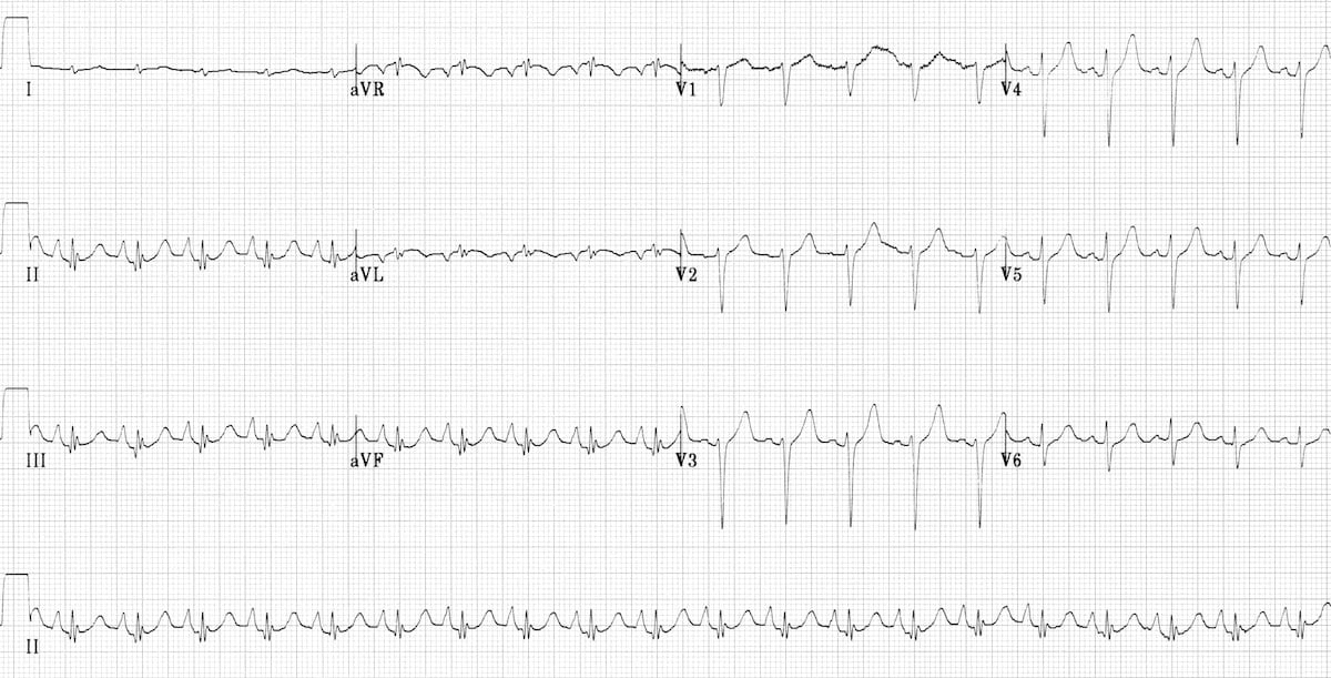
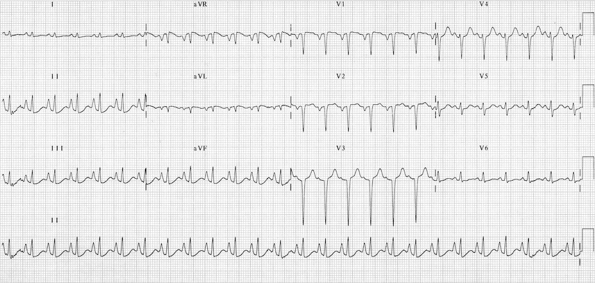
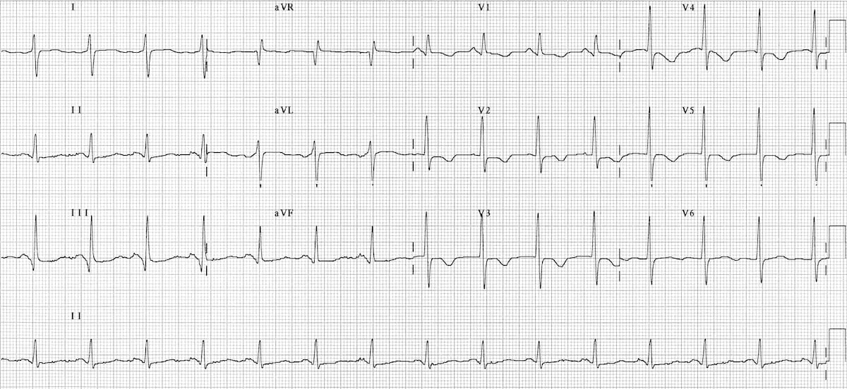
Ekg pulmonary disease pattern. The combination of any of the follow ECG changes suggests increased pulmonary and RV pressures knonw as. S1Q3T3 Pattern of Acute Cor Pulmonale is Classic Pattern also termed as McGinn-White Sign. When CT scans cannot effectively diagnose a pulmonary embolism ECG can be very helpful if there are changes.
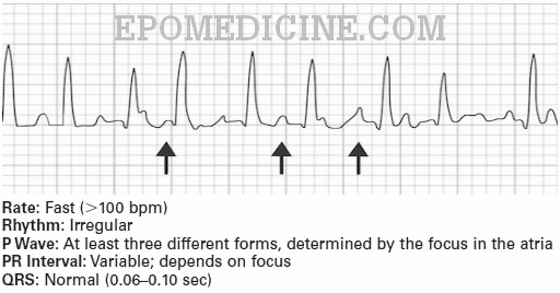
Sinus tachycardia or other types of arrhythmias such as atrial flutter or atrial fibrillation. RBBB complete or incomplete common R ventricular strain T wave inversions R chest V1-4 and inferior leads IIIII aVf Most sensitive up to 99 Classic Change McGinn-White Change S1Q3T3 only 10-20 of PEs. ECG fingdings can be very helpful in diagnosing Pulmonary Embolism.
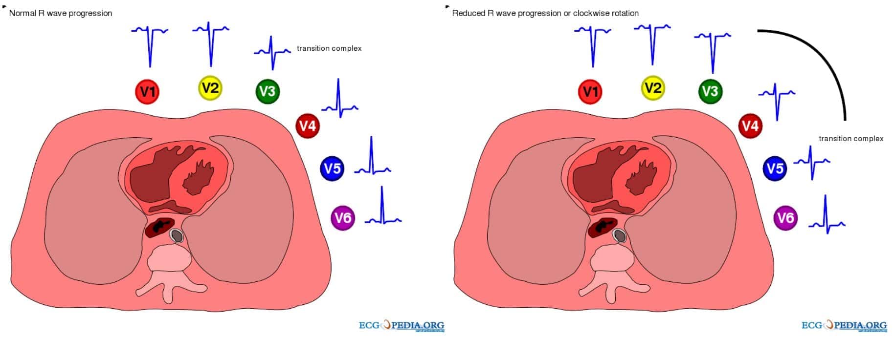
S1Q3T3 Pattern is called classic EKG pattern. Clockwise rotation shift of the RS transition point towards V6 with a persistent S wave in V6 pulmonary disease pattern implying rotation of the heart due to right ventricular dilatation Atrial tachyarrhythmias AF flutter atrial tachycardia 8. Walked on the treadmill and had normal results.
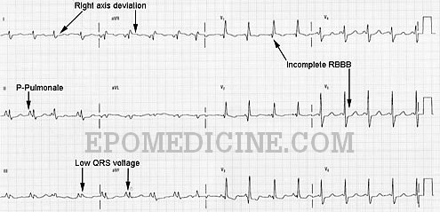
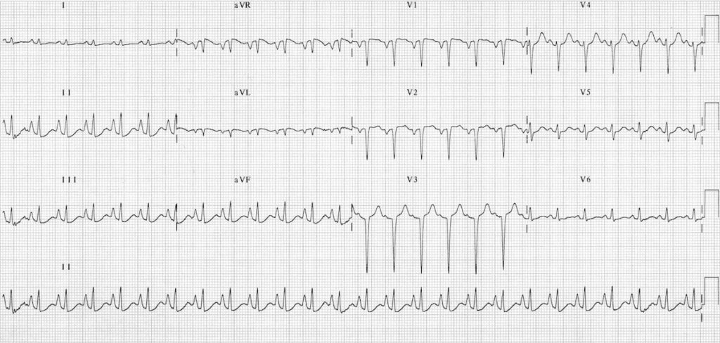
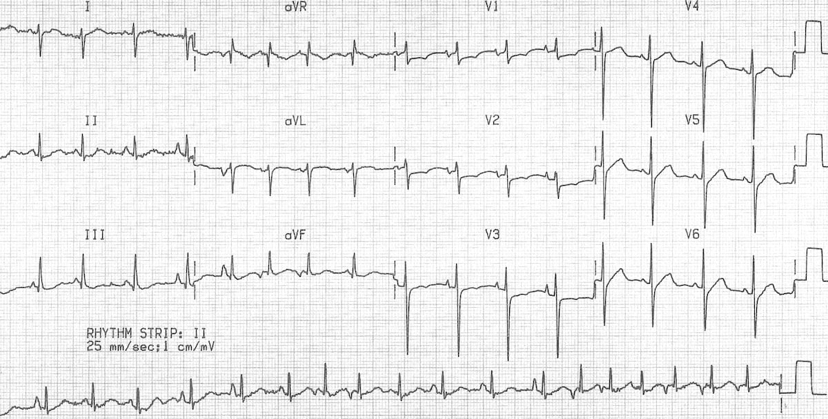
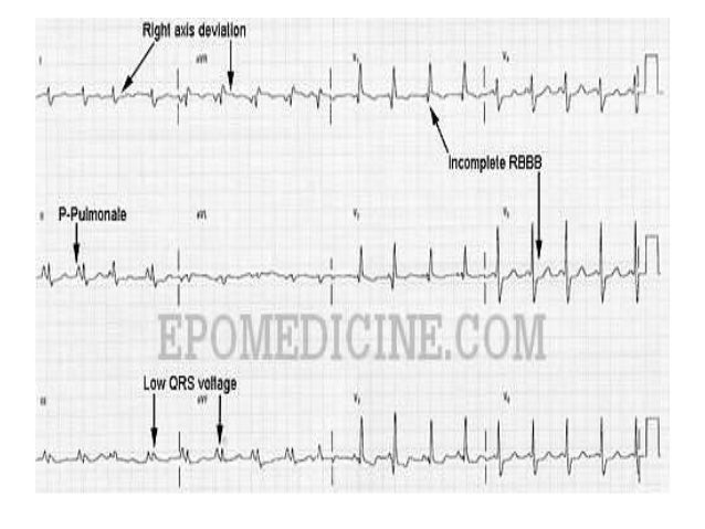
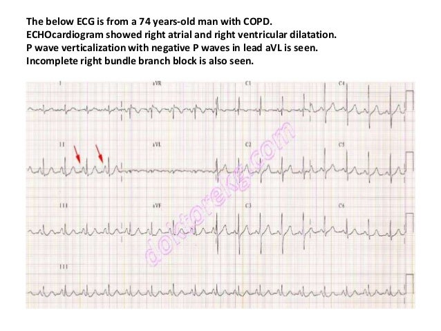
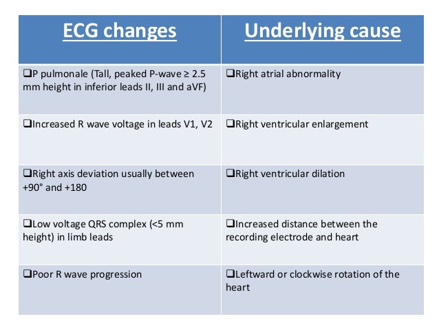
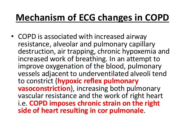
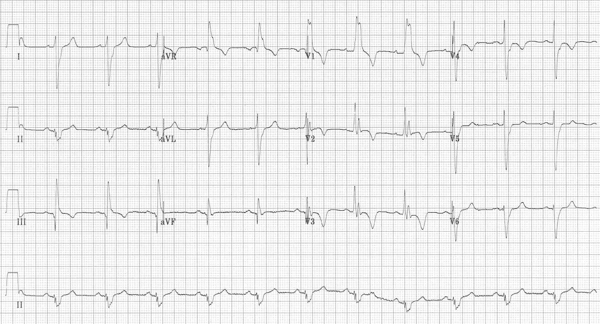
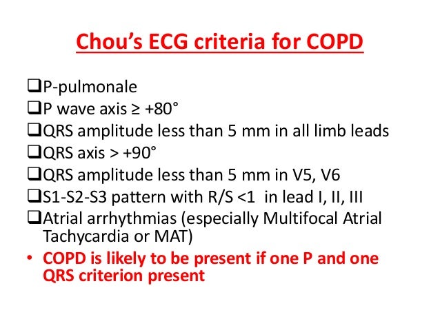
There are various deviations from the normal and no consistent pattern with worsening of airway obstruction1 2 The number of deviations however increases with the Global Initiative for Chronic Obstructive Lung Disease GOLD class3 The most consistent patterns reported have been vertical axes for P and QRS1 4 5 often found in emphysema and increased P-wave amplitude P. The ECG in the Figure was obtained from a 78-year-old man with long-standing pulmonary disease and new-onset heart failure. Suspicion for long-standing pulmonary disease with possible RVHpulmonary hypertension should therefore be raised by the combined ECG findings of rightward axis incomplete RBBB low voltage in several precordial leads and persistent precordial S waves in leads V 4 V 5 V 6 even in the absence of a tall R wave in lead V 1 and ECG criteria for right atrial enlargement.
Tightness in the chest rapid heart beat heart murmur normal pulmonary function test echocardiogram early repolarization in EKG. ECG CHANGES CONSISTENT WITH PULMONARY HYPERTENSION. Most doctors have heard of S1Q3T3.
He also had a normal echocardiogram. Pulmonary Disease Pattern Left anterior fascicular block Left ventricular hypertrophey with QRS widening Non specific T wave abnormality. An R wave 7 mm in V1 or V2.
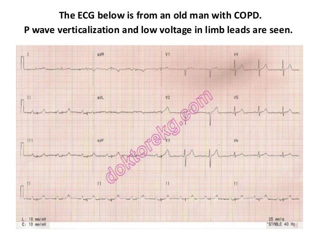
This pattern includes low voltage in the limb leads. Pulmonary embolism on the EKG.
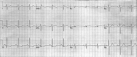
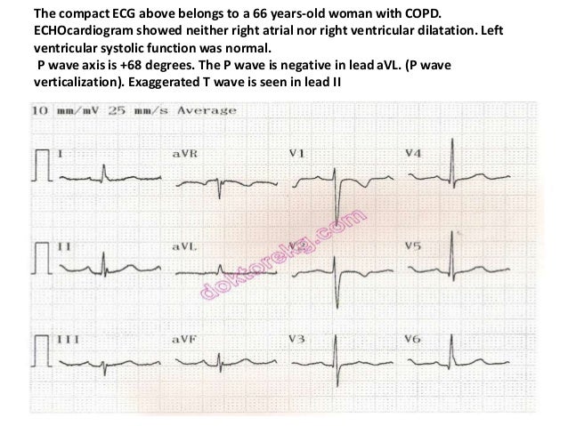
ECG demonstrates many of the features of chronic pulmonary disease.
Based on the low voltage in leads V1 V2 V3 the rightward frontal plane axis incomplete right bundle-branch block and persistent precordial S waves the computer interpreted the overall pattern as consistent with pulmonary disease. Electrocardiography ECG is a useful adjunct to other pulmonary tests because it provides information about the right side of the heart and therefore pulmonary disorders such as chronic pulmonary hypertension and pulmonary embolism. It is also the ECG pattern known to residents and hospitalists. An R wave 7 mm in V1 or V2. Vent rate 69PR int. Walked on the treadmill and had normal results. S1Q3T3 EKG Classic Pattern in Pulmonary Embolism Example. See also Electrocardiography in cardiovascular disorders. Deep S wave in V6 7 mm.
He had a heart murmur as a child but it went away. A rightward P wave axis ie greater than 60 degrees. The combination of any of the follow ECG changes suggests increased pulmonary and RV pressures knonw as. Right bundle branch block S1Q3T3 pattern. ECG demonstrates many of the features of chronic pulmonary disease. RBBB complete or incomplete common R ventricular strain T wave inversions R chest V1-4 and inferior leads IIIII aVf Most sensitive up to 99 Classic Change McGinn-White Change S1Q3T3 only 10-20 of PEs. S1Q3T3 Pattern of Acute Cor Pulmonale is Classic Pattern also termed as McGinn-White Sign.













Post a Comment for "Ekg Pulmonary Disease Pattern"